
New publication (University of Stuttgart), published in GAMM Mitteilungen. The work has been developed within the SFB 1313 research projects B05 and Z02 and the SPP 1897.
"Multi-scale characterization of granular media by in situ laboratory X-ray computed tomography"
Authors
- Matthias Ruf (University of Stuttgart)
- Kianoosh Taghizadeh (University of Stuttgart, SPP 1897)
- Holger Steeb (University of Stuttgart, SFB 1313 research projects B05, C05 and Z02)
Abstract
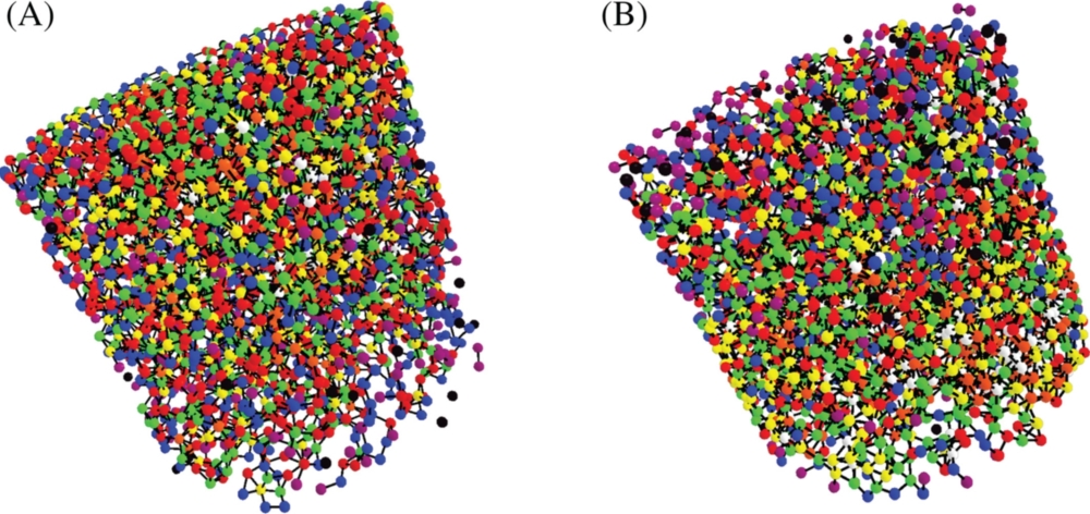
Investigations of biphasic monodisperse soft (rubber) and stiff (glass) particle mixtures under hydrostatic conditions show an interesting behavior with regard to the effective stiffness. P-wave modulus measured by acoustic wave propagation at ultrasonic frequencies showed a significant decline while more soft particles are added, that is, higher rubber volume fractions, due to a change in the microstructure of the granular medium. However, for small volume fractions of soft particles, it could be observed that the P-wave modulus is increasing. This result cannot be explained by classical mixture rules or effective medium theories. For the understanding of those effects, a detailed insight into the microstructure of the granular medium is necessary. To gain this information and link it later back to the measured effective mechanical properties, high-resolution micro X-ray computed tomography (μXRCT) imaging is a well-established tool. With μXRCT imaging, the granular microstructure can be visualized in 3D and characterized subsequently. Combining classical effective characterization methods with μXRCT imaging can help to solve a variety of multi-scale problems. Performing the characterization step in situ, meaning inside the laboratory-based μXRCT scanner, has the advantage that exactly the same samples are mechanically characterized and visualized. To address the mentioned observation above, we designed a low X-ray absorbing oedometer cell with integrated broadband piezoelectric P-wave transducers which enables this kind of investigation inside a laboratory-based μXRCT scanner. The focus of this contribution is on the general experimental methodology which can be transferred to other multi-scale problems. It starts with a description of the image acquisition and ends with the post-processing of the in situ acquired image data. To demonstrate this, cylindrical samples consisting of the same monodisperse rubber and glass particle mixtures that were studied before under hydrostatic stress conditions are considered. Selected results are presented to explain the single steps.

Holger Steeb
Prof. Dr.-Ing.Spokesperson, Project Leader, Research Projects B05, C05, and Z02, Project WIKO, Central Project Z